Introduction
Awareness of the normal ranges for paediatric vital signs is valuable to understand a child’s clinical picture and anticipate the need for escalation. Children’s physiology is different from adults’, and hence, different values are required to determine whether a child falls within or outside the normal range. Paediatric vital sign values are categorised depending on age.1
The Glasgow Coma Scale (GCS) is a widely used scoring system that provides a quick, practical way of assessing consciousness levels in response to various stimuli.2 The GCS was modified to form the PGCS3,4 suitable for use in children, though both are composed of the best response to three parameters: eyes, verbal, and motor.
Vital Signs
Vital signs are a clinical measurement that reflect essential body functions, which include heart rate (HR), respiratory rate (RR), temperature and blood pressure (BP).
Physiology
Children are not simply ‘small adults’ and have different physiology from adults. From neonates to adolescents, children are still physically and cognitively developing. Consequently, paediatric vital signs are categorised according to age groups. The Paediatric Early Warning Sign (PEWS) is a well-adopted tool in many paediatric units to categorise vital signs in children.5 Children are incredibly good at compensating; for example, they can maintain their blood pressure for longer during acute illness; hence, hypotension is a late sign in a sick child.6 For this reason, other organ symptoms and signs can be used to assess circulation in children (such as reduced urine output (UO), mottled/ashen skin, pallor, cool peripheries, and altered mental state). For more detail on this, see the NICE traffic light system for identifying the risk of serious illness in children.7

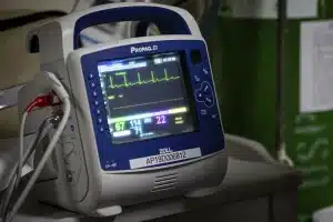
Image 1: A vital signs monitor, used for continuous monitoring of observations when required.
Heart Rate
Children’s resting heart rates are generally higher than adults – the highest values being seen in neonates and decreasing with age. Children have a smaller heart, with a smaller stroke volume and a smaller amount of circulating blood volume than adults, which all increases as they continue to develop.
Cardiac output (CO) is calculated by multiplying heart rate (HR) and stroke volume (SV). This helps explain the higher heart rate seen in younger children. For children to have adequate cardiac output, their heart rate must be higher than in adults.
Respiratory Rate
A child’s respiratory rate will decrease as their lungs develop. There is less space for the gaseous exchange of oxygen and carbon dioxide, hence a higher respiratory rate in younger children.
Normal ranges
The normal values for children can be seen below in table 1.1
| BP (Systolic) | |||||
| Age | RR (breaths per min, 5th-95th centile) | HR (beats per min, 5th-95th centile) | 5th Centile | 50th Centile | 95th Centile |
| Birth | 25-50 | 120-170 | 65-75 | 80-90 | 105 |
| 1m | 25-50 | 120-170 | 65-75 | 80-90 | 105 |
| 3m | 25-45 | 115-160 | 65-75 | 80-90 | 105 |
| 6m | 20-40 | 110-160 | 65-75 | 80-90 | 105 |
| 12m | 20-40 | 110-160 | 70-75 | 85-95 | 105 |
| 18m | 20-35 | 100-155 | 70-75 | 85-95 | 105 |
| 2y | 20-30 | 100-150 | 70-80 | 85-100 | 110 |
| 3y | 20-30 | 90-140 | 70-80 | 85-100 | 110 |
| 4y | 20-30 | 80-135 | 70-80 | 85-100 | 110 |
| 5y | 20-30 | 80-135 | 80-90 | 90-110 | 110-120 |
| 6y | 20-30 | 80-130 | 80-90 | 90-110 | 110-120 |
| 7y | 20-30 | 80-130 | 80-90 | 90-110 | 110-120 |
| 8y | 15-25 | 70-120 | 80-90 | 90-110 | 110-120 |
| 9y | 15-25 | 70-120 | 80-90 | 90-110 | 110-120 |
| 10y | 15-25 | 70-120 | 80-90 | 90-110 | 110-120 |
| 11y | 15-25 | 70-120 | 80-90 | 90-110 | 110-120 |
| 12y | 12-24 | 65-115 | 90-105 | 100-120 | 125-140 |
| 14y | 12-24 | 60-110 | 90-105 | 100-120 | 125-140 |
| Adult | 12-24 | 60-110 | 90-105 | 100-120 | 125-140 |
Table 1: Normal ranges for paediatric vital signs. Adapted from the APLS Aide-Memoires for both boys and girls, from ALSG 6th Edition1
Pathophysiology
There are many reasons why a child’s vital signs could increase or decrease, falling outside of normal ranges.
Heart Rate
Increased: crying, anxiety, dehydration, fever, sepsis, pain, arrhythmias, anaphylaxis, IgE-mediated cow’s milk protein allergy (CMPA), use of medications like salbutamol. Research has shown a linear association between infant pulse rate and body temperature.8
Decreased: normal physiological response (during sleep and in athletes), vasovagal episode, beta-blockers, heart block, hypothyroidism.
Respiratory Rate
Increased: may be caused by lung pathology (e.g. bronchiolitis, pneumonia), metabolic acidosis (e.g. hyperventilation in diabetic ketoacidosis (DKA) or panic attacks), anaphylaxis, and sepsis.9
Decreased: may be caused by central respiratory depression from raised intracranial pressure (ICP), poisoning, hypothermia, encephalopathy, or imminent respiratory arrest.9
Temperature
Temperatures should not be used as a predictor of sepsis,10 however, a temperature <36 or >38.5 is a red flag. However, for historical fever, modalities of temperature measurement should be explored.
Fever is a sign that an immune response is occurring. The level of fever, when interpreted in isolation, does not necessarily correlate to how sick the child is. However, the exceptions to this are:7
- A temperature >390C in infants 3-6 months old
- A temperature >380C in babies <3 months old
Some fevers respond to antipyretics, whilst others don’t. The reason for this is not widely understood; however, we know that a fever is not normally dangerous for a child! Antipyretics can be given to improve symptoms for the child so that they feel better, but do not affect the body’s response to the illness (for example, to a viral infection).
GCS
As mentioned above, the GCS has been adapted for use in children to the PGCS (Paediatric Glasgow Coma Scale). The differences and similarities between the two can be seen below:
| GCS | PGCS | ||
| Behaviour | Response | Score | |
| Eye-opening response | Spontaneously | Spontaneously | 4 |
| To speech | To speech | 3 | |
| To pain | To pain | 2 | |
| No response | No response | 1 | |
| Best verbal response | Oriented to time, place and person | Alert, babbles, coos, words or sentences to usual ability | 5 |
| Confused | Less than usual ability, irritable cry | 4 | |
| Inappropriate words | Cries to pain | 3 | |
| Incomprehensible sounds | Moans to pain | 2 | |
| No response | No response | 1 | |
| Best motor response | Obeys commands | Normal spontaneous movements | 6 |
| Moves to localised pain* | Localizes to supraorbital pain (> 9 months old) or withdraws to touch | 5 | |
| Flexion withdrawal from pain | Withdraws from nailbed pain | 4 | |
| Abnormal flexion (decorticate) | Flexion to supraorbital pain (decorticate) | 3 | |
| Abnormal extension (decerebrate) | Extension to supraorbital pain (decerebrate) | 2 | |
| No response | No response to supraorbital pain (flaccid) | 1 | |
| Total score | Best patient | 15 | |
| Comatose client | 8 or less | ||
| Totally unresponsive | 3 | ||
Table 2: Comparison of Adult GCS and PGCS, adapted from First Aid for free3 and British Paediatric Neurology Association4
*Pain in adults can be elicited by: fingertip pressure, trapezius pinch, or at the supraorbital notch
Any child with GCS <8 will require urgent definitive airway management.
It is worth noting that factors such as alcohol and drugs can ‘mask’ the true level of consciousness, especially in a head injury situation.3
NICE Traffic Light System
The NICE traffic light system for identifying risk of serious illness7 categorises symptoms and signs as high risk (red), intermediate risk (amber), or low risk (green), with recommendations dependent on which category a child falls into.11
Management
If red features suggesting a severe or life-threatening cause of febrile illness are present, the child should be transferred by ambulance to A&E. These could include:
- Features of sepsis or central nervous system (CNS) infection such as bacterial meningitis/meningococcal disease, or encephalitis (see https://cks.nice.org.uk/topics/sepsis/ and https://cks.nice.org.uk/topics/meningitis-bacterial-meningitis-meningococcal-disease/)
- Features of pneumonia or severe dehydration (see https://cks.nice.org.uk/topics/cough-acute-with-chest-signs-in-children/)
If there are non-life-threatening red features, arrange an urgent (within 2 hours) face-to-face assessment (if the child was assessed by telephone) to help guide if hospital admission is necessary.
If there are amber features (with no red features), arrange a face-to-face assessment if the child was initially assessed by telephone or hospital admission, depending on clinical judgement. Consider admission if:
- Infant <3 months with suspected UTI and no alternative focus of infection (to obtain a urine specimen and initiate treatment) (see https://cks.nice.org.uk/topics/urinary-tract-infection-children/)
- No apparent underlying cause for the fever, and the child is unwell for longer than expected for a self-limiting illness
- There is significant parental/carer anxiety and/or difficulty coping due to the family/social situation
If the child can be managed at home, provide advice by one of the following methods, depending on clinical judgement:
- Advice on warning symptoms and signs and when the urgent medical review is needed (see the UK Sepsis Trust leaflet and consider giving to parents to help advise on when to seek further help)
- Arrange a follow-up appointment in primary care
- Liaise with other healthcare professionals, including out-of-hours providers, to ensure direct access for the child if further assessment is required
The child can usually be managed at home if there are green features (with no amber or red features).
If the child can be managed at home:
- Assess for and manage any underlying cause of fever if appropriate
- Consider urine dipstick and urine microscopy and culture if there is unexplained fever and no apparent focus of infection to exclude UTI
- Advise paracetamol or ibuprofen to reduce fever if the child is uncomfortable or distressed, and measures to prevent dehydration (see https://cks.nice.org.uk/topics/feverish-children-management/)
- Advise that routine antipyretics do not reduce or prevent recurrent febrile seizures
- Provide safety netting advice on warning symptoms and signs and when the medical review is needed
References
| No. | Reference |
| 1 | Advanced Life Support Group (ALSG). APLS aide-memoir. APLS 6th Edition ed2015. |
| 2 | Royal College of Physicians an Surgeons of Glasgow. Glasgow Coma Scale [Accessed 10th July 2021]. Available from: https://www.glasgowcomascale.org/. |
| 3 | John Furst. The Glasgow Coma Scale (GCS) for first aiders First Aid for free2013 [updated 18th June 2020Accessed 11th July 2021] |
| 4 | British Paediatic Neurology Association. Child’s Glasgow Coma Scale 2001 [Accessed 11th July 2021]. Available from: https://bpna.org.uk/audit/GCS.PDF. |
| 5 | Lambert V, Matthews A, MacDonell R, Fitzsimons J. Paediatric early warning systems for detecting and responding to clinical deterioration in children: a systematic review. BMJ Open. 2017;7(3):e014497. |
| 6 | Hagedoorn NN, Zachariasse JM, Moll HA. Association between hypotension and serious illness in the emergency department: an observational study. Archives of Disease in Childhood. 2020;105(6):545. |
| 7 | National Institute for Health and Care Excellence. Traffic light system for identifying risk of serious illness 2019 [Accessed 11th July 2021]. Available from: https://www.nice.org.uk/guidance/ng143/resources/support-for-education-and-learning-educational-resource-traffic-light-table-pdf-6960664333. |
| 8 | Hanna CM, Greenes DS. How much tachycardia in infants can be attributed to fever? Annals of Emergency Medicine. 2004;43(6):699-705. |
| 9 | Friend DA, Mann DJ. Approach to the Seriously Unwell Child TeachMePaediatrics [updated 10th August 2018Accessed 10th July 2021]. Available from: https://teachmepaediatrics.com/emergency/emergency-medicine/approach-to-the-seriously-unwell-child/ |
| 10 | National Institute for Health and Care Excellence. Sepsis: recognition, diagnosis and early management, NICE guideline [NG51] 2016 [updated 13 September 2017Accessed 10 March 2021] |
| 11 | National Institute for Health and Care Excellence. Scenario: Feverish children – risk assessment 2020 [Accessed 11th July 2021]. Available from: https://cks.nice.org.uk/topics/feverish-children-risk-assessment/management/feverish-children-risk-assessment/#the-nice-traffic-light-system. |
| 12 | The UK Sepsis Trust. Sam’s Story [Accessed 11th July 2021]. Available from: https://sepsistrust.org/about/about-sepsis/patient-stories/sam/. |
