Introduction
Acute mastoiditis is an intratemporal complication of otitis media. It occurs due to otitis media infection spreading to involve the bone of the mastoid air cells directly.
This article will discuss the epidemiology, pathophysiology, clinical features, diagnosis, management and complications of acute mastoiditis.
Epidemiology
Around 75% of cases of acute mastoiditis occur in children under the age of 2 with a peak incidence at age 6-13 months. Since the introduction of antibiotics, acute and chronic mastoiditis have become quite rare. The incidence in developed countries is 1.2-6.1 cases per 100,000.
Pathophysiology
The mastoid air cells are a collection of air-filled spaces located in the mastoid process of the temporal bone. They communicate with the middle ear via a small canal known as the aditus to mastoid antrum.
Infection can spread from the middle ear into the mastoid air cells causing infection of the bone of the mastoid air cells. This in turn leads to breakdown of the fine trabeculae of the mastoid air cells along with collection of pus in the mastoid antrum. As the pus is enclosed under pressure, this causes localised bone necrosis, which can spread to form a sub-periosteal abscess.
The abscess can usually be found in 1 of 3 places:
- Behind the pinna in an area known as Macewen’s triangle, or higher
- Superior to the pinna towards the zygomatic process
- Over the squamous temporal bone
Risk Factors
- More common in young children
- Immunocompromised patients
- Pre-existence of cholesteatoma
Clinical Features
The diagnosis of acute mastoiditis is based mainly on thorough history-taking and clinical examination. Supporting investigations may be undertaken.
As acute mastoiditis is a complication of otitis media, a history of acute or recurrent otitis media is one of the clinical features. In addition, there may be a history of:
- Otalgia
- Blocked ear or deafness
- Pyrexia (can be swinging)
- Infants may present with irritability, excessive crying and feeding problems
On examining a patient with acute mastoiditis, one may find:
- Unwell child who may be lethargic
- Red bulging eardrum
- Alternatively, ear discharge with a perforated ear drum
- Oedema of posterior and superior part of deep ear canal
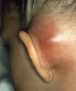
- Tenderness behind the pinna – MacEwen’s triangle
- Pinna can be pushed forwards by swelling and appear more prominent
Neurological examination is usually normal; any abnormal findings may suggest advanced disease:
- Abducens nerve (CN VI) palsy
- Facial nerve (CN VII) palsy
- Facial pain due to involvement of CN Va (ophthalmic branch of trigeminal nerve)
Differential Diagnoses
- Infected pre-auricular sinus (located near the front of the ear)
- Infected/inflamed post-aural lymph node
- Langerhans cell histiocytosis
- Rhabdomyosarcoma (malaise with mastoid swelling + radiological opacity)
Investigations
Supporting investigations may be undertaken to aid in diagnosis of mastoiditis but should not delay in administration of treatment.
Laboratory tests that may be helpful include:
- Ear swab – if discharging ear or post-aural abscess is oozing; sent for MC+S
- Blood tests – raised inflammatory markers including WCC and CRP
CT head and mastoid with contrast is indicated in all cases of mastoiditis apart from cases of simple uncomplicated mastoiditis. CT images will typically show coalescence of the mastoid air cells along with an opaque mastoid and middle ear. It is also helpful in excluding intracranial complications of mastoiditis.
MRI head under sedation or anaesthesia is helpful in cases of suspected complicated mastoiditis due to its increased sensitivity in highlighting soft tissue collections or vascular problems. Magnetic resonance venogram (MRV) is best at identifying venous thrombosis, which can be a complication of mastoiditis.
Management
The initial management of acute mastoiditis is intravenous antibiotics as an inpatient. The choice of antibiotic will be based on local guidelines but should cover likely causative organisms including Streptococci and Staphylococcus aureus; high-dose co-amoxiclav or ceftriaxone are usually chosen. An oral switch can be considered if the child is recovering and pyrexia has settled. It is recommended that oral antibiotics should be continued for a further 14 days.
Indications for surgical intervention:
- Uncomplicated mastoiditis that fails to improve clinically after 48 hours of intravenous antibiotics
- Continuing pyrexia, malaise
- Persistent erythema or soft tissue swelling behind the ear in mastoid region
- Presence of complications of mastoiditis
Different techniques may be used to treat surgically depending on difference in practice including:
- Needle aspiration of pus
- Incision and drainage
- Cortical mastoidectomy to formally open mastoid antrum and drain the infection
During the procedure, a myringotomy and drainage of middle ear pus may also be undertaken.
Complications and Prognosis
Since the introduction of antibiotics, the number of patients with complications associated with mastoiditis has greatly reduced.
Complications of mastoiditis can mainly be separated into 2 categories: extracranial and intracranial complications.
Extracranial:
- Facial nerve palsy
- Hearing loss – conductive and sensorineural
- Labyrinthitis
- Subperiosteal abscess
- Cranial osteomyelitis
Intracranial:
- Intracranial infections including meningitis; epidural, temporal lobe or cerebral abscess; subdural empyema
- Dural sinus thrombosis
The prognosis for the majority of cases that are diagnosed early is excellent with a low likelihood of complications occurring.
References
| [1] | Chesney, J., Black, A. and Choo, D. (2013). What is the best practice for acute mastoiditis in children?. The Laryngoscope, [online] 124(5), pp.1057-1058. Available at: https://onlinelibrary.wiley.com/doi/abs/10.1002/lary.24306 |
| [2] | Hussain, S. and Turner, A. (2015). Logan Turner’s Diseases of the ear, nose and throat. 11th ed. CRC Press. |
| [3] | Lin, H.W., Shargorodsky, J., & Gopen, Q. Clinical Strategies for the Management of Acute Mastoiditis in the Pediatric Population. Clinical Pediatrics. Vol 49, Issue 2, pp. 110 – 115. |
| [4] | Loh, R., Phua, M., & Shaw, C. (2018). Management of paediatric acute mastoiditis: Systematic review. The Journal of Laryngology & Otology, 132(2), 96-104. doi:10.1017/S0022215117001840 |

