Introduction
Gastroschisis is a full-thickness abdominal wall defect in which fetal abdominal organs protrude outside the abdomen with no protective membrane covering them. Direct intestinal exposure to amniotic fluid in utero leads to chemical reactions, creating a thick inflammatory film or peel over the bowel.1
Epidemiology
Gastroschisis has a reported incidence of 1-6 per 10,000 live births.2 There is a 10 to 16-fold higher incidence for women under 20 years to have a child with gastroschisis than women aged 25-29.1
Pathophysiology
The exact mechanism of gastroschisis is currently unknown.3 However, suggested theories include compromised vascular supply to the anterior abdominal wall, a defect in the primordial umbilical ring, or abnormal involution of the right umbilical vein creating a weakened point at risk of rupture.1,2,4
Risk Factors
Many potential agents have been implicated, but no exact relationship has been established:1,5
- Maternal smoking (possibly due to placental insufficiency and abnormal development of the vascular system)
- Maternal age <20 years old
- Environmental exposures e.g. Nitrosamines
- Maternal cyclooxygenase inhibitors use e.g. aspirin and ibuprofen
Clinical Features
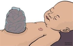
Gastroschisis can present either visible at birth or detected early on prenatal ultrasound scans at 20 weeks.4 The abdominal organs are herniated outside the abdominal cavity through a full-thickness opening, often found to the right of the umbilical cord.2 Commonly involved organs include the small intestines, large intestines, liver and stomach. A distinguishing feature of gastroschisis from omphalocele is that no membrane covers the abdominal contents (Figure 1).
Other possible clinical features include intestines which appear swollen, inflamed, thickened, and/or short. A thick fibrous peel can be seen over the contents secondary to inflammation from amniotic fluid exposure. Also, the neonatal abdominal cavity may appear smaller than expected.2
Gastroschisis is associated with intestinal malrotation, and 10-15% of cases are associated with intestinal atresia.4 This may be due to the small size of the abdominal wall defect, causing constriction of mesenteric blood supply and ischemia of the exposed bowels. Extra-intestinal or structural abnormalities are not commonly associated with gastroschisis, unlike omphalocele.2 Additionally, most infants are affected by malabsorption and hypomotility, leading to delayed bowel function.4

Figure 1- The clinical features of gastroschisis. Adapted from the CDC ‘Facts about grastroschisis’.
Differential Diagnosis
Omphalocele – abdominal wall defect presenting with membranous sac covering over abdominal contents, frequently associated with other congenital abnormalities.2
Cloacal Exstrophy – abdominal wall defect where a segment of the large bowel and two halves of the bladder is present outside the abdominal cavity.1
Physiological gut herniation – herniation of the bowel in early pregnancy at 6-8 weeks and subsequent return into the abdominal cavity at 12-13 weeks in-utero.2
Investigations
Gastroschisis is a mainly clinical diagnosis.1 It can be diagnosed by prenatal ultrasound or upon birth through physical examination.4
Laboratory Tests
Alpha-fetoprotein is routinely measured in antenatal screening and typically be elevated in abdominal wall defects.6 This may result from direct protein loss from the intestine into the surrounding amniotic fluid. Elevated alpha-fetoprotein indicates further assessment through antenatal ultrasound.1
Imaging
Gastroschisis can be picked up on routine antenatal ultrasounds in the second trimester when bowel outside the abdominal cavity can be detected.1 Ultrasonography can show echogenic and dilated loops of bowel freely floating in the amniotic cavity. Coloured doppler may help localise the herniation in relation to the umbilical cord.2 Intestinal atresia may be detected in some cases.
After birth, infants are not routinely given any further investigations unless there are signs of dysmorphia. Although, families are routinely offered genetic counselling to aid in planning for future children.1
Management
Earlier detection through antenatal ultrasound scans is helpful for discussion and planning with families about their birth plans. Women with gastroschisis babies can have normal vaginal deliveries unless a cesarean section is indicated for other reasons. Most women are induced around 37 weeks at a tertiary care centre for immediate neonatal and surgical care.3,4
Immediate Management
Following delivery, post-natal management includes immediate fluid resuscitation and maintaining adequate temperature. A sterile, clear covering over the herniated contents protects the bowel, preventing evaporation, heat loss, and infection. The infant is placed on his right side to prevent kinking of mesenteric vessels.1
Definitive Management
Surgery aims to reduce the protruding organs and close the abdominal wall defect. Larger defects may need a staged surgical approach to return the contents into the abdominal cavity gradually; this will involve placing the bowel in a clear sac called a silo, which is tightened until there is enough space to reduce the bowel completely (Figure 2). This prevents further damage to the gut due to tight space within the abdomen.3
Following surgery, a nasogastric tube is inserted to decompress the bowel, and parenteral feeding is commenced while the inflammatory peel recovers and the bowel starts functioning.1
Complications
Complications include:1,2
- Abdominal Compartmental Syndrome
- Persistent bowel dysfunction
- Wound infection
- Necrotizing Enterocolitis
- Short gut syndrome
- Abdominal Compartmental Syndrome
Prognosis
Gastroschisis has reported survival rates from 87-100%. However, patients may require extended hospital stays. Intestinal atresia is the most important prognostic factor for morbidity in gastroschisis.1
References
| No. | Reference |
| 1 | Leaphart, CL. Omphalocele and gastroschisis. https://bestpractice.bmj.com/topics/en-gb/1158 (accessed March 2021). |
| 2 | Rasuli B, Radswiki, et al.. Gastroschisis. https://radiopaedia.org/articles/gastroschisis?lang=gb (accessed March 2021). |
| 3 | The Department of Specialist Neonatal and Paediatric Surgery. Gastroschisis. https://www.gosh.nhs.uk/conditions-and-treatments/conditions-we-treat/gastroschisis/ (accessed March 2021). |
| 4 | National Organization for Rare Disorders. Gastroschisis. https://rarediseases.org/rare-diseases/gastroschisis/ (accessed March 2021). |
| 5 | Centers for Disease Control and Prevention. Facts about Gastroschisis. https://www.cdc.gov/ncbddd/birthdefects/gastroschisis.html (accessed March 2021). |
| 6 | Fahrion C. Gastroschisis. https://surgery.ucsf.edu/conditions–procedures/gastroschisis.aspx (accessed March 2021). |

