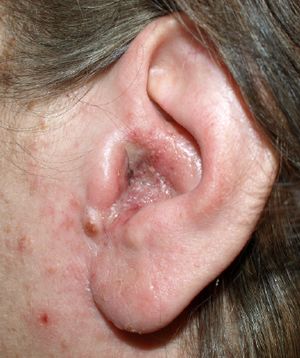
Otitis externa is an inflammation of the external ear canal and can be either acute or chronic in nature. Acute otitis externa lasts less than 3 weeks whereas chronic otitis externa lasts more than 3 months.
Otitis externa can also be classified by the area which it affects; localized otitis externa is an infection of a hair follicle in the ear which can develop into a boil. Diffuse otitis externa is widespread inflammation of the skin and subdermis.
Malignant otitis externa arises when the infection spreads to the mastoid and temporal bones causing osteomyelitis (1). The term malignant was adopted due to the poor prognosis associated with this outcome, and should not be confused with a neoplastic condition.
Epidemiology
Otitis externa is common and each year more than 1% of the UK population will develop it (2). The incidence usually increases towards the end of summer due to warmer temperatures and more people participating in swimming (2). It is slightly more common in women than men (1).
Pathophysiology
Otitis externa is an infection of the skin in the external auditory canal (3).
There are different causes of otitis externa (1):
- Bacterial infection – most commonly Pseudomonas Aeruginosa or Staphylococcus Aureus. The bacteria usually enter the ear after 1 of 4 events:
- Blockage of the canal
- Absence of cerumen due to excess cleaning
- Trauma
- Alteration of pH within the canal (3).
- Fungal infection
Risk Factors
- Hot and humid climates
- Swimming
- Older age
- Diabetes Mellitus
- Narrowing/obstruction of the auditory canal
- Over-cleaning leading to a lack of wax in the canal
- Wax build-up
- Eczema
- Trauma
- Radiotherapy to the ear (4)
Clinical Features
From history
The main symptoms are (4):
- Pain
- Itching
- Discharge
- Hearing loss
From examination
Otoscopy may show the following features:
- Oedema
- Erythema
- Exudate
- Mobile tympanic membrane
Other features may include:
- Pain on movement of tragus or auricle
- Pre-auricular lymphadenopathy (4)
Differential Diagnosis
This presentation could also be due to (1,5):
- Acute otitis media with perforation of the TM
- Furunculosis (infection of a hair follicle in the cartilaginous part of the ear canal)
- Viral infections
- Tumours of external auditory canal
- Cholesteatoma
- Foreign body
- Impacted wax
- Skin conditions eg. acne, psoriasis, contact dermatitis, seborrhoeic dermatitis
Investigations
Generally, investigations are not needed for otitis externa. However, if treatment has failed or the infection is thought to be with atypical bacteria or fungus, swabs of the ear can be sent off for microscopy and culture (4).
Management
General advice (6):
- Avoid getting the ear wet use a cap for showering and swimming
- Remove any discharge by gently using cotton wool, DO NOT put cotton buds into the ear
- Remove any hearing aids and earrings
- Use painkillers – paracetamol and ibuprofen
Specific management:
- Antibiotic or antifungal ear drops are generally the mainstay of treatment (6). A pope wick can be used to get the drops into the ear if the canal is closed.
- If there is cellulitis or lymphadenopathy then oral antibiotics are indicated (4).
- In cases of chronic otitis externa, acetic acid and corticosteroid ear drops are used (4).
Complications
The condition can be complicated by (7):
- Abscesses
- Stenosis of the ear canal due to a build-up of thick, dry skin
- Perforated ear drum
- Cellulitis
- Malignant otitis externa – infection spreads to mastoid and temporal bones
Prognosis
Most cases of otitis externa resolve within a few days of starting treatment. Only two subsets of patients require follow up; those who may have an underlying cholesteatoma and those who have systemic disease predisposing to persistent symptoms (4).
References
| (1) | NICE Clinical Knowledge Summaries, “Otitis Externa”, December 2016. [Online]. Available: https://cks.nice.org.uk/otitis-externa#!topicsummary [Accessed August 2017]. |
| (2) | BMJ Best Practice, “Otitis Externa”, April 2017. [Online]. Available: http://bestpractice.bmj.com/best-practice/monograph/40/basics/epidemiology.html [Accessed August 2017]. |
| (3) | A Waitzman, “Otitis Externa”, May 2017. [Online]. Available: http://emedicine.medscape.com/article/994550-overview#a4 [Accessed August 2017]. |
| (4) | “Otitis Externa and Painful, Discharging Ears”. [Online]. Available: https://patient.info/doctor/otitis-externa-and-painful-discharging-ears [Accessed August 2017]. |
| (5) | “Otitis Externa Differential Diagnosis”, April 2017. [Online]. Available: http://bestpractice.bmj.com/best-practice/monograph/40/diagnosis/differential.html [Accessed August 2017]. |
| (6) | NHS Choices, “Otitis Externa”, October 2015. [Online]. Available: http://www.nhs.uk/Conditions/Otitis-externa/Pages/Treatment.aspx [Accessed August 2017]. |
| (7) | NHS Choices, “Otitis Externa – complications”, October 2015. [Online]. Available: http://www.nhs.uk/Conditions/Otitis-externa/Pages/Treatment.aspx [Accessed August 2017]. |

