Introduction
Epidemiology
Primary bone cancers are rare, making up only 0.2% of all cancers diagnosed annually in the UK. However, they account for approximately 5% of all European childhood cancers diagnosed annually, and osteosarcoma is the most common childhood primary bone cancer.1
Osteosarcoma most commonly occurs in teens and young adults, with a second peak in presentation in adults over 50. It is more common in males than females and commonly affects Afro-Caribbean patients in adolescence.2, 3
Pathophysiology
The cause is largely unknown; however, a widely accepted theory is that DNA mutations occur in rapidly dividing osteoblasts, such as during pubertal growth spurts, leading to malignant transformation. This theory is based on osteosarcomas commonly occurring at the time of pubertal growth spurts and in locations of fast bone growth.1,2,4,5 Osteosarcomas most commonly occur in the limbs, at the metaphysis of long bones such as the femur and tibia.1,5
There are three significant types of osteosarcoma- osteoblastic, chondroblastic and fibroblastic. The type depends on how well-differentiated the cells are when the oncogenic event occurs. The osteoblastic subtype arises from the most highly differentiated cells, the fibroblastic subtype arises from the least differentiated cells, and the chondroblastic subtype is somewhere in between.2,5
Risk factors
There are very few known risk factors of osteosarcoma in childhood, although there is an increased risk in individuals who are taller than average.5,6 Other than this association, some known genetic conditions are known to predispose osteosarcoma. These include:
- Li-Fraumeni syndrome- an autosomal dominant germline mutation affecting the p53 protein (a tumour suppressor protein).
- RB1 mutation- this affects the retinoblastoma protein, causing hereditary retinoblastoma. Osteosarcoma is the most common tumour caused by this mutation, aside from retinoblastoma itself.
- Other genetic conditions include Rothmund-Thomson Syndrome, Bloom Syndrome, Werner Syndrome, Rapadilino, and Diamond-Blackfan Anaemia.
These genetic conditions only account for a small number of osteosarcoma cases.5
Clinical features
The most common feature of osteosarcoma is pain, usually at the tumour site, although some individuals will experience referred pain. It is often intermittent, worsens at night, and is resistant to analgesia.1,2,5,7
Many individuals will feel or see a lump, which is sometimes warm to touch and tender on palpation. The absence of a lump does not rule out osteosarcoma.5,7
Some people will present with mobility issues, most commonly stiffness. If the tumour is in the leg, it can cause a limp or affect the ability to walk. Some individuals experience non-specific symptoms, such as fatigue, weight loss, and headache.5,7
Clinical examination may reveal only tenderness at the tumour site, or there may be a lump. The absence of these features does not rule out a primary bone tumour; if there is clinical suspicion, the individual should be referred for imaging, even if their examination is normal.
Red flag features are bone pain that occurs at night and/or increases in intensity. If these symptoms are present, it must be assumed that a bone tumour is present until proven otherwise by investigation.
Differential diagnosis
Differential diagnoses include:
- Secondary bone tumours (this is more likely in adults than in children)
- Benign bone tumours
- Infection (e.g. osteomyelitis)
- Developmental abnormalities including fibrous or cystic defects
- Trauma
- Metabolic bone disease (e.g. osteomalacia)
- Haematological malignancy
Investigations
If osteosarcoma or a primary bone tumour is suspected, blood tests should be checked and radiographs of the affected area should be taken in two planes.1,2,5
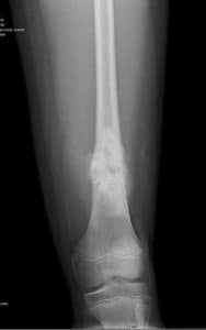
Features that may be present on radiographs include:
- Bone destruction
- New bone formation
- Periosteal swelling
- Soft tissue swelling
It is important to note that a normal radiograph does not rule out osteosarcoma. Clinical suspicion with persistent bone pain or night pain needs urgent MRI and referral to a sarcoma centre.

Figure 1- An X-Ray showing osteosarcoma of the distal tibia ©radiopedia.org
Blood tests should include FBC, U&E, CRP, ESR, bone profile and lactate dehydrogenase. None of these are diagnostic for osteosarcoma but may help differentiate between other possible diagnoses, offer some prognostic value, and may be helpful as baseline blood tests for potential treatment such as chemotherapy.1
Following plain radiographs, staging tests such as CT, MRI, or isotope bone scans should be performed.
To get a definitive diagnosis, a biopsy of the affected area is required; ideally, this should take place at a specialist sarcoma centre, where any future surgery will be undertaken. Patients should also be referred to relevant paediatric bone sarcoma MDT.1
Management
Management of osteosarcoma usually involves surgery and chemotherapy.
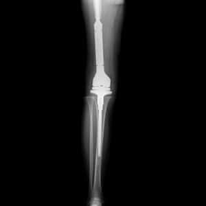
The aim of surgery is usually to remove the whole tumour with sufficient margins. There may be the option for limb-salvage surgery, which involves reconstruction of the limb. Alternatively, amputation may be required. Provided there has been no metastasis, the aim of surgical removal of the tumour is usually curative.
Due to the aggressive nature of osteosarcoma and the chance of micrometastatic disease, chemotherapy is part of the standard treatment. The regimen has remained essentially unchanged since the 1970s and commonly involves neoadjuvant chemotherapy before surgery, followed by adjuvant chemotherapy.5 Commencing chemotherapy before surgery has no long-term prognostic benefit. Still, it helps to improve symptoms rapidly, may help with the treatment of micrometastatic disease and allows time for the customised endoprosthesis to be made. There is also the benefit that histological response to the chemotherapy can be seen on removing the tumour.5
First-line chemotherapy in patients aged 30 or under involves doxorubicin, cisplatin and high-dose methotrexate +/- mifamurtide (an immunotherapy agent). This treatment is the same for local and metastatic diseases.1
Complications and prognosis
The prognosis for osteosarcoma is notoriously poor compared to the majority of cancers. Five-year survival of childhood osteosarcoma is approximately 60% compared to the 80% five-year survival of all childhood cancers.8 Bone cancers are one of the leading causes of cancer deaths amongst teens and young adults.9 This may be attributable to late diagnosis and the aggressive nature of osteosarcoma, with its ability to micro-metastasise.
Other complications include the risk of pathological fractures and metastatic disease. Many children and young adults will have metastatic disease at diagnosis, drastically worsening prognosis.5,7
References
| No. | Reference |
| 1 | Gerrand C, Athanasou N, Brennan B, Grimer R, Judson I, Morland B et al. UK guidelines for the management of bone sarcomas. Clinical Sarcoma Research. 2016;6(1). |
| 2 | Deyrup A, Siegal G. Practical orthopedic pathology. Philadelphia: Elselvier Inc; 2016. |
| 3 | National Cancer Intelligence Network. Bone Cancer Incidence and Survival. Birmingham; 2012. |
| 4 | Valery P, Laversanne M, Bray F. Bone cancer incidence by morphological subtype: a global assessment. Cancer Causes & Control. 2015;26(8):1127-1139. |
| 5 | Tattersall L, Davison Z, Gartland A. Osteosarcoma. Encyclopedia of Bone Biology. 2020;:362-378. |
| 6 | Mirabello L, Pfeiffer R, Murphy G, et al. Height at diagnosis and birth-weight as risk factors for osteosarcoma. Cancer Causes Control. 2011;22(6):899-908. |
| 7 | Bone Cancer Research Trust. 2020 Patient Survey. Leeds; 2020. |
| 8 | Public Health England. Childhood Cancer Statistics, England Annual report 2018. London: PHE Publications; 2018. |
| 9 | Public Health England. 13-24 year olds with cancer in England Incidence, mortality and survival. London: PHE Publications; 2018. |

